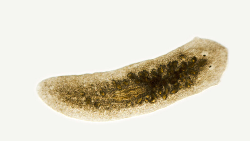
Photomicrograph of planaria, a flatworm, Dugesia species. Slight motion blur in head. Freshwater stream, San Luis Obispo, California. Live specimen. Wet mount, 2.5X objective, transmitted brightfield illumination.
To regenerate a head, you first have to know where your tail is – Ars Technica
2025-04-18T18:40:04Z
Planaria can’t replace a missing head until after the tail develops sufficiently.
For those of us whose memory of high school biology hasn’t faded entirely, planarians will probably sound very familiar. They’re generally used as an example of one of the extreme ends of regenerative capacity. While some animals like mammals have a limited ability to regenerate lost tissues, planarians can be cut roughly in half and regenerate either an entire head or entire tail, depending on which part of the body you choose to keep track of.
In doing so, they have to re-establish something that is typically only needed early in an animal’s development: a signaling system that helps tell cells where the animal’s head and tail are. Now, a US-based team asked a question that I’d never have thought of: What happens if you cut the animal in half early in development, while the developmental head-to-tail signaling system is still active? The answer turned out to be surprisingly complex.
Heads or tails?
Planarians are small flatworms that would probably be living quiet lives somewhere if biologists hadn’t discovered their ability to regenerate lots of adult tissues when damaged. The process has been well-studied by this point and involves the formation of a cluster of stem cells, called a blastema, at the site of damage. From there, many of the signals that control the formation of specialized tissues in the embryo get re-activated, directing the stem cells down the developmental pathways needed to reproduce any lost organs.
Critical to this process is a signaling system that helps inform the animal of where its head is. Signaling molecules called wnts help developing embryos form what’s called the anterior-posterior axis by telling the animal where its posterior resides. If it’s needed, wnt signaling may also be reactivated during regeneration after the loss of major portions of the animal, inducing the blastema to form new tail structures. That reactivation takes place in the animal’s muscle, which appears to be a key tissue in the process of sensing the damage and inducing regeneration.
The question being asked here is what happens if the damage that induces regeneration happens while the animal is still developing at a time when wnt signaling is normally patterning the animal’s organs. To find out, the researchers started taking planarian embryos (from the species named Schmidtea polychroa) and slicing them in half.
Auto-posted from news source